Summary
In the fall of 2015, Dysautonomia International was pleased to provide a $15,000 grant from the POTS Research Fund to study Vasomotor Neuropathy in POTS to Professor Roy Freeman, Professor of Neurology at Harvard Medical School and Director of the Center for Autonomic and Peripheral Nerve Disorders at Beth Israel Deaconess Medical Center in Boston.
Approximately 50% of postural orthostatic tachycardia syndrome (POTS) patients have a reduction in small fiber nerve density, most often in the lower limbs. This is described as "neuropathic POTS." In these patients, POTS symptoms are believed to occur due to damage to the peripheral nerves that control blood vessel function (vasomotor nerves). Dr. Freeman's lab recently developed a novel technique to measure vasomotor nerves via skin biopsy. The vasomotor technique will complement the lab's previously reported work on the sensory nerves in the skin in patients with POTS and will provide a direct measure of the pathology that underlies neuropathic POTS.
This technique is different than the skin biopsies already available for small fiber sensory and sudomotor nerve innervation (which Dr. Freeman's lab previously developed). The vasomotor skin biopsy technique also differs from the Quantitative Sudomotor Axon Reflex Test (QSART or Q-Sweat), a non-invasive test which is often used to evaluate sudomotor neuropathy in POTS patients. Sudomotor nerves innervate the sweat glands - sudo is Latin for sweat. While sudomotor dennervation in POTS is helpful in identifying a neuropathic process in some POTS patients, loss of sweat gland function is not responsible for the majority of POTS symptoms, whereas diminished or absent control of blood vessel tone, regulated by vasomotor nerves, is very relevant to the symptoms that POTS patients experience.
Additional Information
Professor Freeman provided the following explanation of the Vasomotor Neuropathy in POTS Study.
Postural orthostatic tachycardia syndrome is a heterogeneous disorder characterized by symptoms of orthostatic intolerance and an excessive increase in heart rate upon attaining the upright position. A number of different pathophysiologic mechanisms underlie this disorder, and several subgroups of patients with POTS have been reported with distinct but overlapping pathophysiologies. Among these, is postural tachycardia that is secondary to damage to the peripheral nerves innervating the blood vessels, also known as neuropathic POTS.
We have demonstrated that individuals with neuropathic POTS have reduced intra-epidermal nerve sensory nerve fiber density compared to control subjects. However, to date, a reduction in the neurovascular innervation of individuals with POTS has not been demonstrated.
To quantify neurovascular innervation, we have developed several novel measurement techniques. Skin biopsies, taken from the distal leg and distal thigh, were obtained from healthy individuals and individuals with peripheral nerve disease. Biopsies are stained with protein gene product 9.5 (PGP 9.5) and CD31 (a vascular endothelial marker) and imaged by confocal microscopy.
The density of capillaries, the subepidermal capillary plexus, and the deeper dermal capillary densities are quantified using a novel unbiased stereologic counting method. In addition, the nerve fiber densities within the sub-epidermal plexus and the deeper dermal plexus are quantified. Finally, the co-localized neuro-vascular innervation in the sub-epidermal plexus and the deeper dermal vasculature were quantified.
The development of these novel quantitative techniques required extensive testing and validation. At this time, we have successfully standardized these quantitative techniques. We believe that this technique will be of value in understanding the pathological process that underlies neuropathic POTS. | | 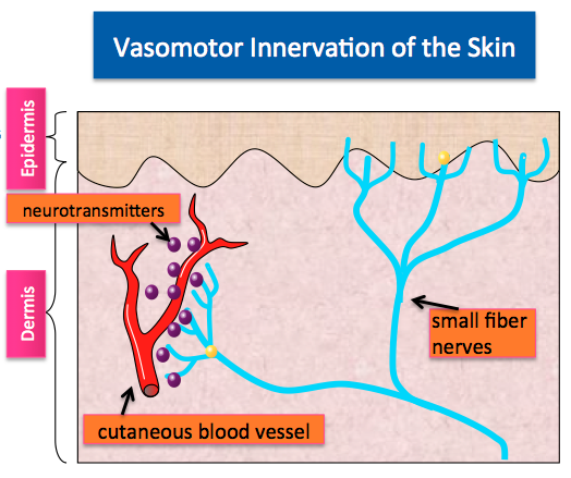 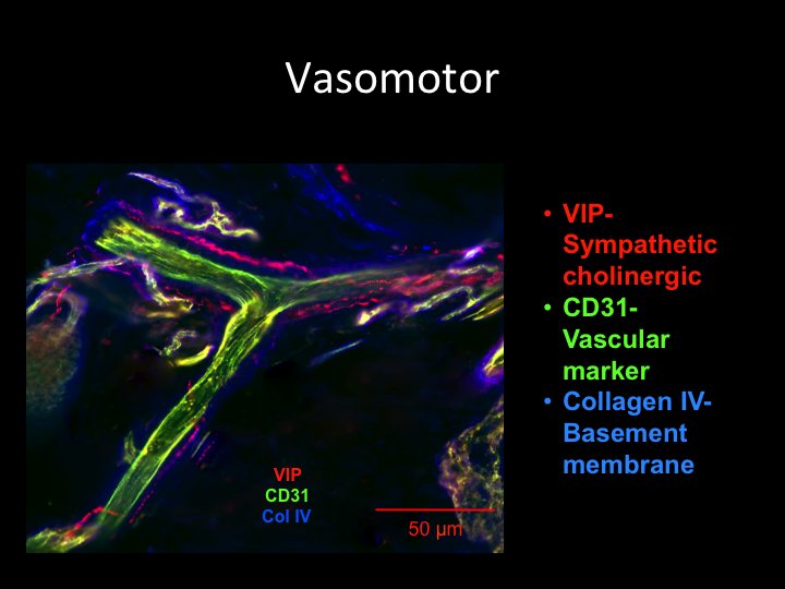
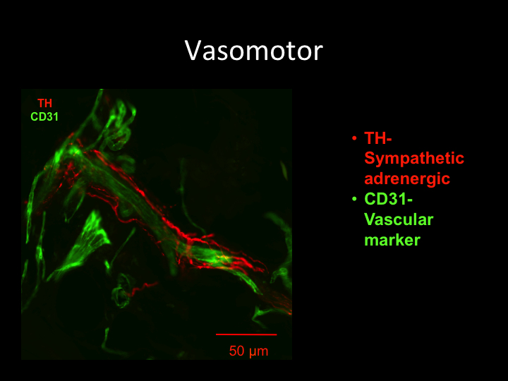
Top image adapted from Prof. Freeman's diagram. Bottom two images are stained vasomotor nerves from Dr. Freeman's lab. Blood vessels are in green, vasomotor (autonomic) nerves are in red and collagen is in blue. |